Quiz Challenge December(ii)
Case Study: Asymptomatic Elderly Lady on Routine Fundal Exam
Dr Daniel Chiu
Diagnoses ?
Click for answer.
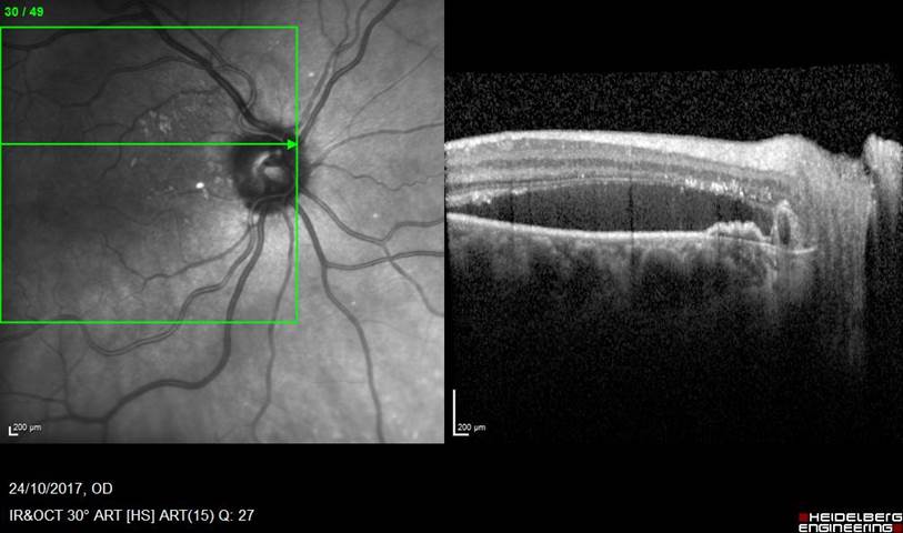
Answer: Right peripapillary choroidal polyp with lipid exudate
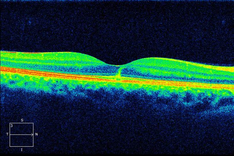
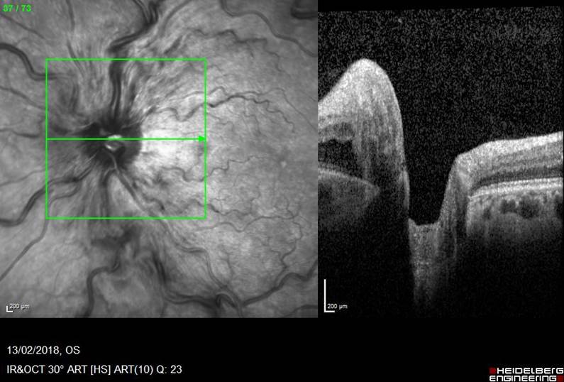
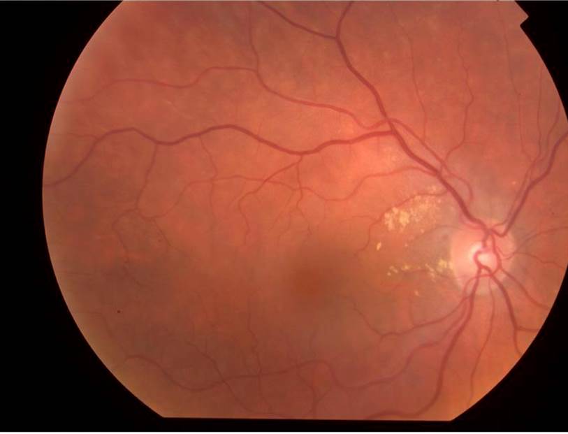
Comment: Reddish small lesion at about 10 o’clock position at disc margin can be visible in the colour photo. OCT across that area show roundish elevation budding out of the RPE layer and associated surround RPE irregular elevation is common. Subretinal fluid accumulation and chronic lipid deposit are also common features. Peripapillary choroidal neovascular membrane is a possibility but usually there will be some haemorrhage outlining the neovascular membrane or some more fibrosis visible