The Price Of Beauty
Case Study: The Price of Beauty
Dr Janice Thean
With the increasing use of dermal fillers, we are starting to see some devastating but rare complications resulting in partial or complete visual loss. Of the documented cases, improvement of visual acuity in patients with vascular occlusion after filler injection is extremely rare, and there has been no complete recovery of vision after initial injury. Loss of vision tends to be instantaneous at the time of dermal filler injection. Often, hyalase is injected on the spot to rectify the condition with limited success.
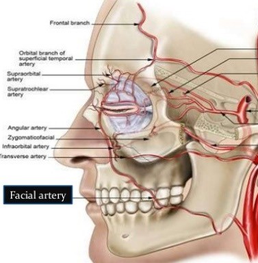
Loss of vision from filler can occur following an injection anywhere on the face, not just around the eye area. Areas including around the nasolabial fold, the lips can all cause blindness. Basically, if the filler is accidentally injected into any part of the facial artery, it can travel along the artery all the way into the retina. Apart from visual loss, injecting filler into the facial artery can cause soft tissue damage / necrosis, destroying / killing certain parts of the face i.e. nasal cartilage.
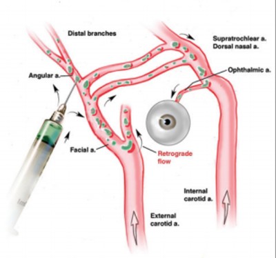
It is thought that this complication is a result of retrograde flow. This occurs when an injection is made under a higher force, the injected substance enters the external carotid vessel at a high enough force that it flows backward into the internal carotid and into the eye.
Case
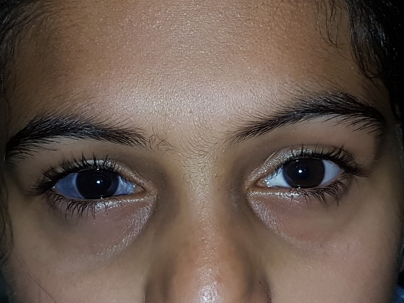
A 28 year old woman presented with immediate blurred vision following dermal fillers to her right cheek. When the visual changes occurred, her doctor immediately proceeded to treat her with hyalase. Unfortunately, there was no recovery of her VA. Apart from visual impairment, the right side of her face was red and swollen for 2 weeks.
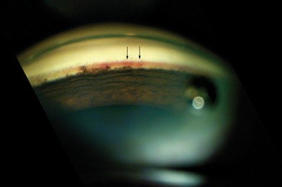
Presenting visual acuity was of 6/12 on the right and 6/6 on the left unaided. Amsler chart revealed a small right inferior scotoma. Examination of the fundus was normal with no obvious signs of emboli or retinal ischemia. The only clue was seen on OCT angiography where there was a small window defect noted just superior to the fovea. Baseline VF was normal.
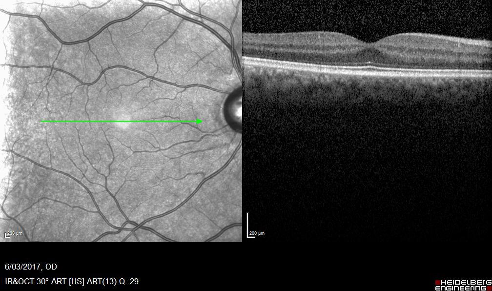
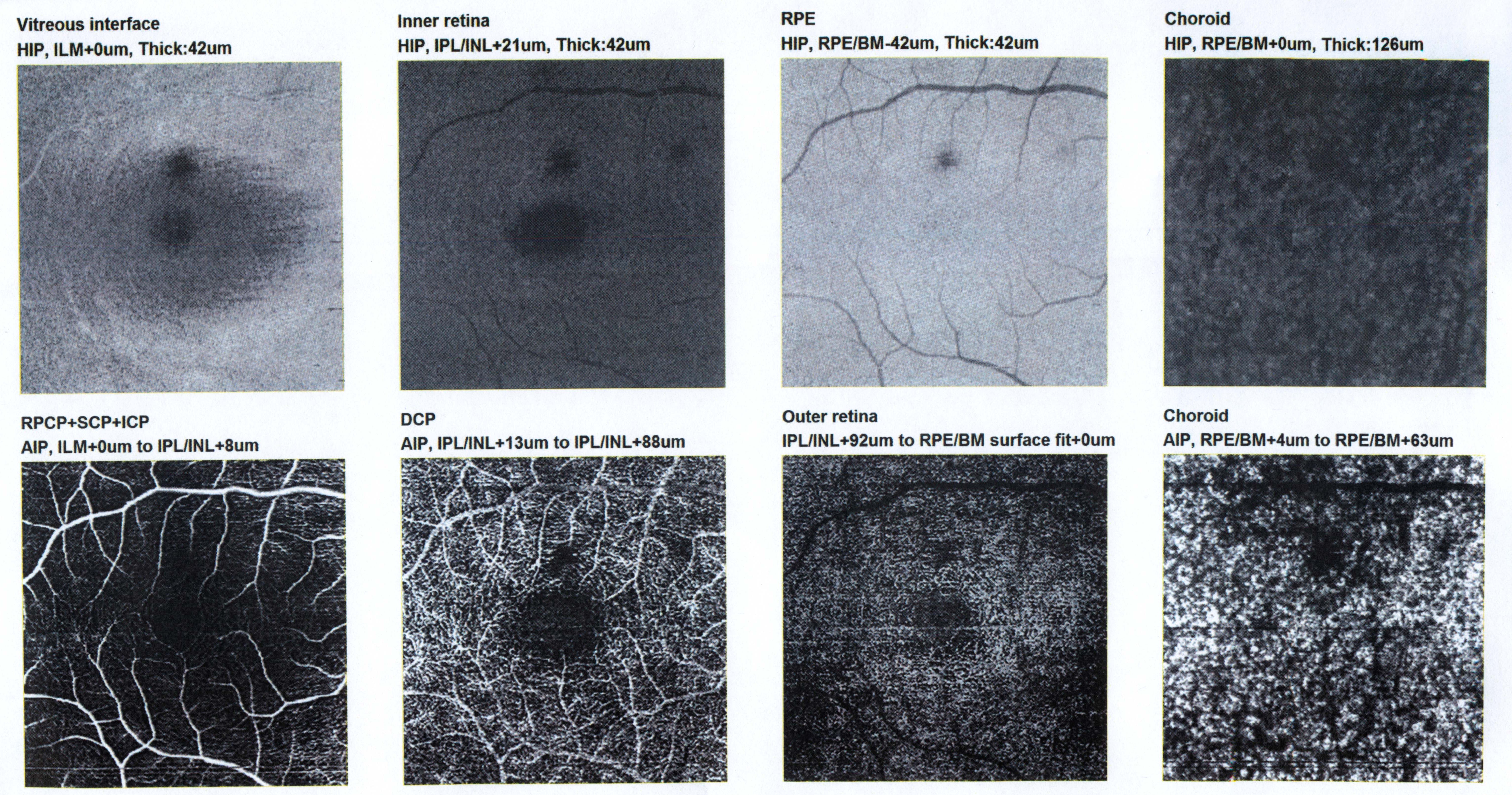 Figure 1: OCT angiogram on presentation
Figure 1: OCT angiogram on presentation
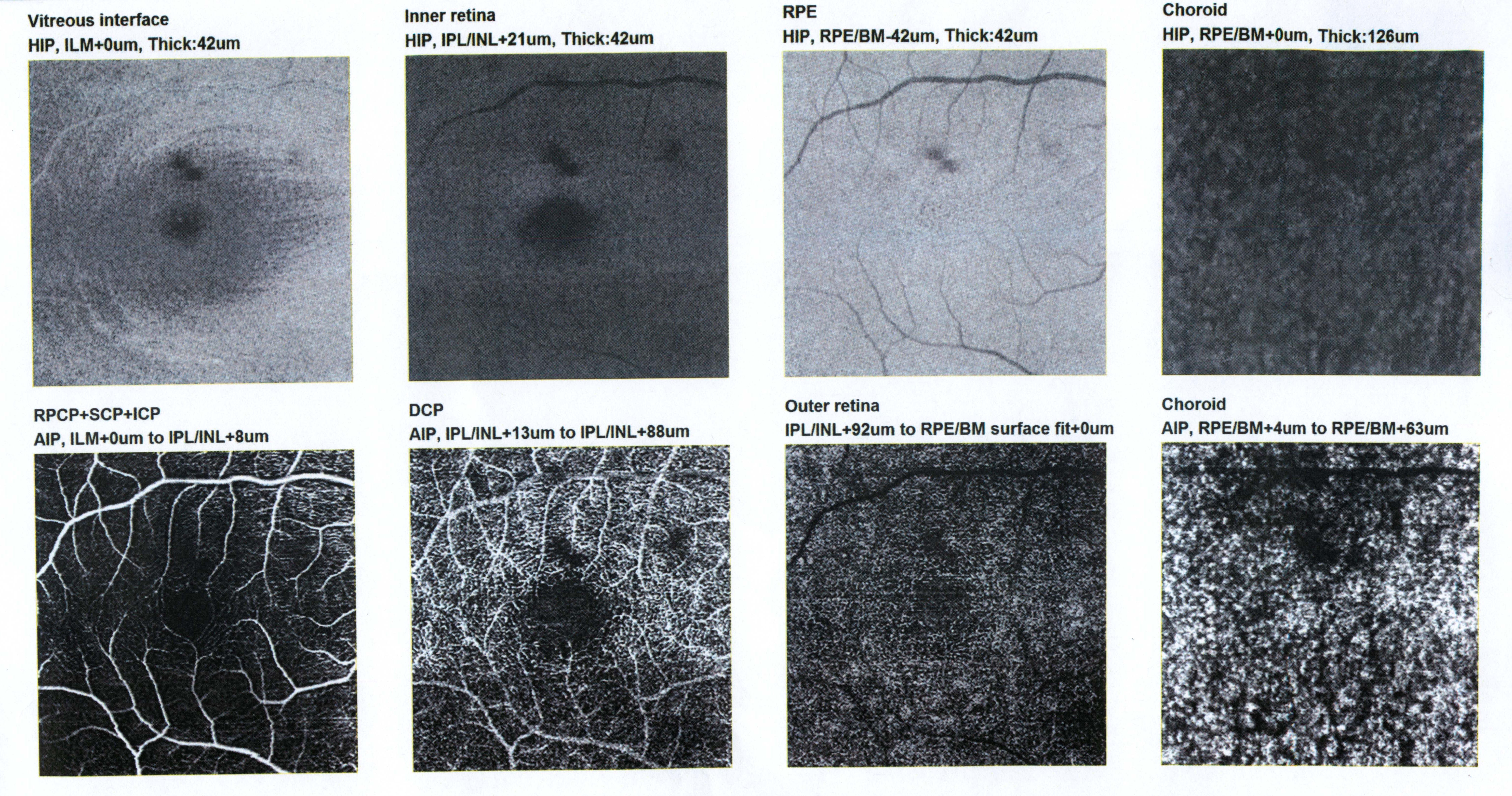 Figure 2: OCT angiogram 2 months later showing no improvement.
Figure 2: OCT angiogram 2 months later showing no improvement.